BLUF:
- Recognize HyperK or the potential for HyperK
- ABCs, monitor, EKG!!! Repeat lab if suspecting iatrogenic hemolysis.
- If EKG changes are present OR potassium>6.0…initiate the treatment below.
- Stabilize the Myocardium
- Calcium Gluconate (10%): 10ml vial IV = 1g of CaGl. Administer via PIV, OR
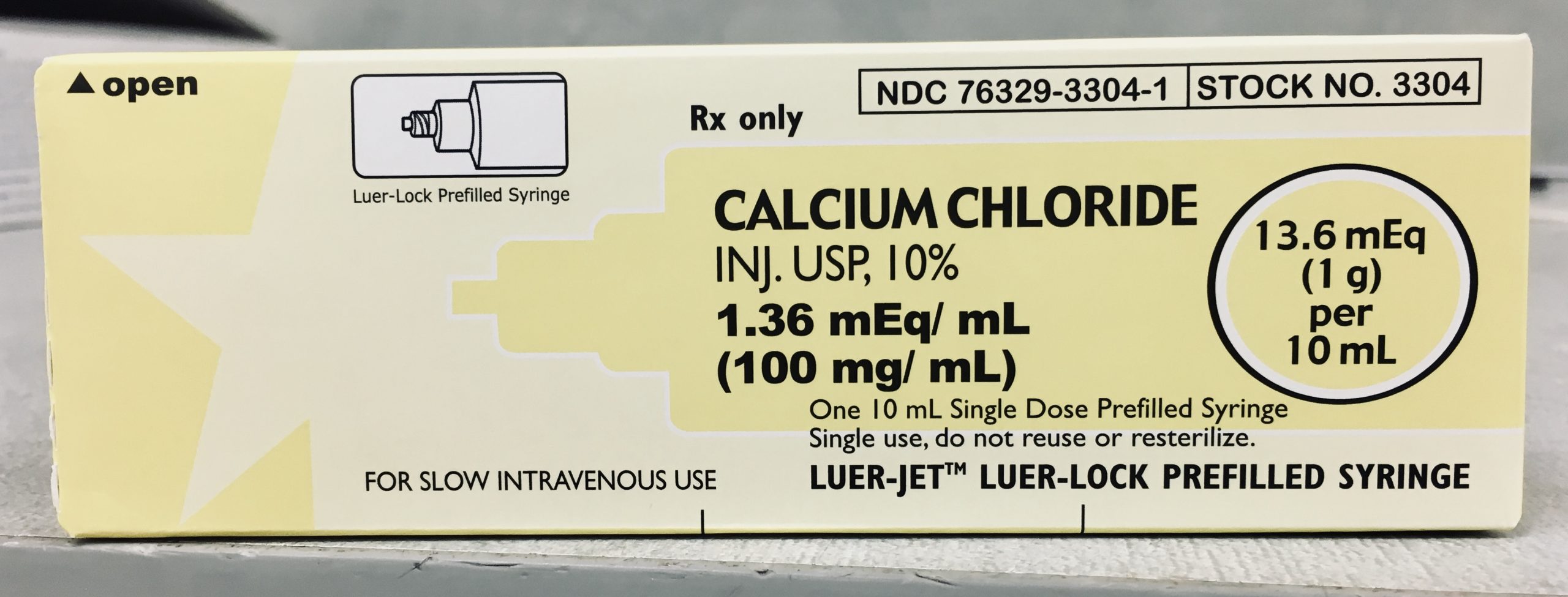
- Calcium Chloride (10%): 10ml ampule IV = 1g of CaCl. Only administer via CVC due to risk of extravasation injury, or administer emergently via PIV during cardiac arrests.
- Push K Intracellularly
- Albuterol: 10-20mg nebulized “high-dose” albuterol. Repeat as needed.
- Insulin/Glucose: 5-10 units of insulin per 1 ampule of D50 IV if normoglycemic. If hyperglycemic, hold D50. In ESRD, decrease insulin dose.
- Bicarbonate: 1 ampule = 50mEq NaHCO3. Only consider if acidemic, or administer empirically in crashing patient. Repeat as needed.
- Eliminate K from the body
- IVF: Increases renal excretion of K. Use NS or LR. LR does not worsen hyperK.
- Lasix: 40-80mg IV. If still making urine.
- Dialysis: The ultimate correction for hyperK in renal failure. Temporize with medications until dialysis available.


IN-BRIEF:

CAUSES:
– Pseudohyperkalemia. Hemolyzed blood sample, most common cause of hyperK.
– CKD/ESRD. Often missed dialysis.
– Acidosis.
– Rhabdomyolysis. Myocytes lyse spilling K into the serum.
– Adrenal Insufficiency. Classic triad of hypotension, hyperK, hypoNa.
– Medications. K sparing diuretics, ACEIs, ARBs.
SERUM LEVELS:
– 5.0-6.0 = “Mild” Hyperkalemia
– 6.1-7.0 = “Moderate” Hyperkalemia
– >7.0 = “Severe” Hyperkalemia
“CLASSIC” EKG CHANGES:
1. T-waves peak
2. PR lengthens, P-waves flatten
3. QRS widens, P-waves disappear
4. Sine-wave appears
5. Ventricular fibrillation, asystole, death


BACKGROUND:
Physiology. Hyperkalemia causes two separate clinical effects. First, excessive extracellular potassium reduces intracellular potassium efflux making the intracellular environment more positive and the cell membrane potential less electronegative. This increases cardiac myocyte excitability increasing the risk of arrhythmias. Second, worsening hyperkalemia “poisons” sodium channels worsening their function and slowing action potential conduction velocities leading to the widening intervals seen on EKGs.

Calcium. Calcium stabilizes the myocardium nearly immediately by effectively reversing the relative effects of hyperK on the cardiac myocyte. Calcium makes the “threshold potential” less negative, decreasing cardiac myocyte excitability. Calcium also restores the original electrochemical gradient of the cardiac myocyte membrane, thus increasing action potential conduction velocities and re-narrowing EKG intervals.

- There are two different types of calcium that can be given – Calcium Gluconate (CaGl) and Calcium Chloride (CaCl).
- CaGl is stored in 10ml vials in the Pyxis and can be given via PIV. Each vial contains 1 gram of CaGl providing 4.65mEq of elemental calcium.
- CaCl is stored in 10ml prepackaged ampules in the code cart and should not be given via PIV except during codes due to the risk of extravasation injury. Each ampule contains 1 gram of CaCl providing 13.6mEq of elemental calcium.
- One gram of CaCl contains three times the amount of elemental calcium as one gram of CaGl due to the much larger molecular mass of CaGl, so give CaCl in emergent situations due to its higher potency.

EKGs. The classically taught “linear progression” of EKG changes in hyperkalemia is not necessarily true and does not necessarily correlate with specific serum potassium levels. Nearly half of patients hyperkalemic to values of ~7.0 will show no EKG changes or EKG changes that are not specific to hyperkalemia. Each patient’s electrophysiologic response to hyperkalemia is different, and arrhythmias can occur at any time without warning. So practically, treat hyperkalemia aggressively with calcium if there are any EKG changes or if the serum potassium value is greater than 6.0.
Albuterol. Albuterol shifts K intracellularly by stimulating the Na-K ATPase pump, it acts immediately, and lowers K by up to 1mEq/L. The studies showing this benefit used high doses of nebulized albuterol (10-20mg), so dose accordingly. Albuterol’s effects are synergistic with insulin.
Insulin. Insulin also shifts K intracellularly by stimulating the Na-K ATPase pump, it exerts its effects within 20 minutes, and lowers K by up to 1mEq/L. Give 10 units of insulin per 1 ampule of D50 (25g of dextrose in 50ml water) if normoglycemic. If hyperglycemic, hold the D50. If renal failure is present, give lower doses of insulin since insulin is renally cleared (ex. 5 units per 1 ampule of D50, etc).
Bicarbonate. Sodium bicarbonate use in hyperkalemia is controversial, studies have shown little benefit. But while throwing the kitchen sink at a crashing hyperkalemic patient, then include some bicarbonate, especially if profoundly acidotic or if there are other indications such as a TCA toxicity. It just may help.
Lasix. “Furosemide” eliminates potassium from the body by inhibiting the Na-K-Cl cotransporter in the ascending limb of the loop of Henle causing Na, K, and Cl to be excreted in the urine. It exerts its diuretic effects within five minutes when given IV, and begins the definitive correction of hyperkalemia. The patient must still make urine though, which may not be the case in renal failure.
Intravenous Fluids. IV Fluids also work in tandem with the renal excretion of potassium, and is synergistic with potassium wasting diuretics. Either NS or LR can be used – LR does not worsen hyperkalemia.
Dialysis. Dialysis provides rapid correction of hyperkalemia, especially in end-stage renal disease, but it is costly and requires significant logistical preparation. Dialysis can be arranged while temporizing hyperkalemia with other treatment modalities.
Resins. Ion exchange resins – “Kayexalate,” given PO or PR – trade sodium for potassium in the colon and eliminate potassium in the stool. Since these resins are dependent upon bowel movements and exert their effects over days, they play no role in the emergent management of hyperkalemia. They have also been associated with intestinal necrosis in some cases.
RRWCT. Regular “Really” Wide Complex Tachycardias. Amal Mattu recommends that when a RWCT that is “really” wide – >200ms – is recognized, then consider and treat metabolic (hyperkalemia) or toxicologic (TCA toxicity, other sodium channel blockers) causes with empiric sodium bicarbonate and calcium. He considers it a “clean kill” if you treat a “RRWCT” with a sodium channel blocker (amiodarone, procainamide, lidocaine) as ACLS recommends. RRWCTs are likely due to hyperkalemia or TCA toxicity, and sodium channel blockers will promptly kill these patients.
INTEL
- Tintinalli, Judith E., et al. Tintinalli’s Emergency Medicine: A Comprehensive Study Guide. Eighth edition. Fluids and Electrolytes, Chapter 17, p. 101-106. New York: McGraw-Hill Education, 2016.
- Hilton J et al. Management of Hyperkalemia in the ED. EMResident. 2018; 45(4): issue 4: 22-23. Online article.
- Parham WA et al. Hyperkalemia Revisited. Tex Heart Inst J. 2006; 33(1): 40–47. PMC1413606.
- Farkas J. Myth-busting: Lactated Ringers is safe in hyperkalemia, and is superior to NS. Emcrit.org. 2014. Link to Article.
- Ashurst J et al. Evidence-Based Management Of Potassium Disorders In The Emergency Department. Emerg Med Pract. 2016 Nov 22;18. PMID: 27775507.


Leave a Reply